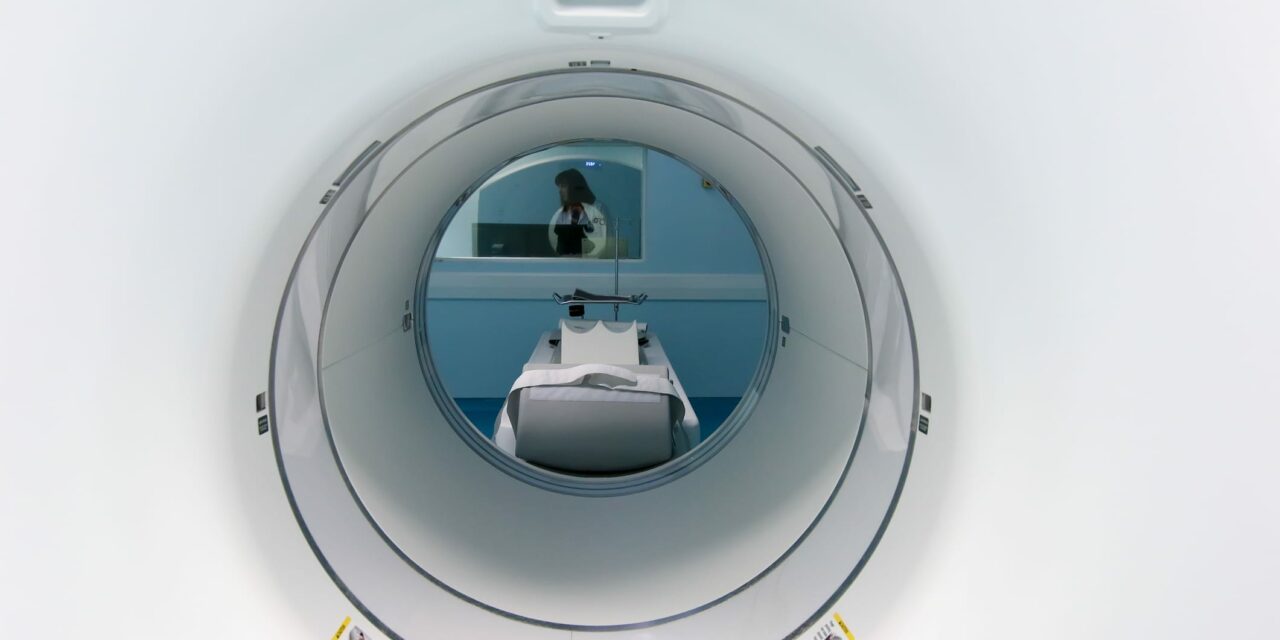
Positron Emission Tomography (PET) imaging has significantly evolved, providing critical advancements in the early detection of various cancers. These innovations are enhancing the accuracy, specificity, and sensitivity of cancer diagnosis, enabling earlier and more precise interventions. Here’s how recent developments in PET imaging are shaping the future of oncology.
Innovative Radiotracers
One of the most notable advancements in PET imaging is the development of novel radiotracers. Traditional tracers like ^18F-FDG have been widely used, but new tracers are now providing more detailed and specific information about tumor biology. For example, the development of fibroblast activation protein inhibitor (FAPI) tracers has shown improved detection capabilities, especially in differentiating malignant from benign lesions in breast and gynecologic cancers. FAPI tracers can detect tumors that might not be visible with standard ^18F-FDG PET/CT, enhancing early diagnosis and treatment planning (Frontiers) (Stanford Medicine).
Enhancing Sensitivity and Resolution
Recent advancements in total-body PET systems have dramatically improved the detection sensitivity and spatial resolution of PET scans. These systems allow for more comprehensive imaging of the entire body, capturing detailed metabolic activity in a single scan. This improvement is crucial for detecting small or early-stage tumors that might be missed with less sensitive imaging techniques. Studies have shown that these advanced PET systems can identify tumor heterogeneity and early metabolic changes, providing a more accurate assessment of the cancer’s extent and progression (Nature).
Real-Time Tumor Tracking
Innovative PET technologies are also enhancing the precision of cancer treatment through real-time tumor tracking. For instance, Stanford Medicine is pioneering a novel approach known as biology-guided radiation therapy (SCINTIX), which integrates PET imaging with radiotherapy. This technology uses PET tracers that bind to cancer cells, providing real-time feedback on tumor location and activity. The radiation delivery system adjusts dynamically based on the tracer signals, allowing for more precise targeting of moving tumors or multiple metastases. This method aims to minimize side effects and improve treatment efficacy by accurately targeting cancer cells while sparing healthy tissue (Stanford Medicine).
Preclinical and Clinical Applications
Recent preclinical studies have demonstrated the potential of advanced PET tracers in improving cancer detection and monitoring. For example, novel PET tracers like ^68Ga-DOTA-CCK-66 have shown promise in detecting medullary thyroid cancer lesions with higher accuracy. These tracers bind specifically to cancer cells, providing clear and detailed images that can guide targeted therapies. Ongoing clinical trials are further evaluating these tracers to confirm their efficacy and potential for broader clinical application (ScienceDaily).
Conclusion
The advancements in PET imaging are revolutionizing early cancer detection and management. Novel radiotracers, improved imaging systems, and innovative real-time tracking technologies are enhancing the precision and accuracy of cancer diagnosis. These developments not only facilitate earlier detection of malignancies but also support more personalized and effective treatment plans. As research continues, the integration of these advanced PET technologies in clinical practice promises to significantly improve patient outcomes and the overall effectiveness of cancer care.
References
- “Advances in PET Imaging of Cancer,” Nature Reviews Cancer.
- “Applications of FAPI PET/CT in the Diagnosis and Treatment of Breast and Gynecologic Malignancies,” Frontiers. Accessed
- “Stanford Medicine First to Try Out Novel Tumor-Targeting Radiation Therapy Machine,” Stanford Medicine News Center. “Novel PET Tracer Enhances Lesion Detection in Medullary Thyroid Cancer,” ScienceDaily.


